
Virus Budding

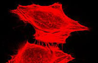
Retrovirus particles entering cells. HIV particles carrying the VSV G envelope were engineered to incorporate high levels of Green Fluorescent Protein (GFP) [first slide]. Chinese hamster ovary cells were infected with highly concentrated preparations of virus and fixed for imaging 2 hours later. The microtubule network has been stained red with rhodamine-coupled antibodies [second slide]. Virus cores are readily detected on the cell surface and within the cytoplasm.
Transferrin Receptor

Aberrant intracellular trafficking of the transferrin receptor in a clone of mutant Rat2 cells isolated after selection for resistance to retrovirus infection. The internalization of the transferrin receptor can be visualized by the location of the red stain after exposure of the cells to rhodamine-labelled transferrin. The receptor is internalized more slowly and remains more diffuse than in wild-type cells. The microtubule network has been stained green.
VSV1
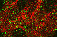
Retrovirus particles entering cells. HIV particles carrying the VSV G envelope were engineered to incorporate high levels of Green Fluorescent Protein (GFP). Chinese hamster ovary cells were infected with highly concentrated preparations of virus and fixed for imaging 2 hours later. The microtubule network has been stained red with rhodamine-coupled antibodies. The virus cores are readily detected on the cell surface and within the cytoplasm.